
Scleroderma vs Morphea: Identifying Skin Changes After 40
Learn to distinguish between localized morphea and systemic scleroderma in your 40s. Explore symptoms, the role of estrogen, and the essential diagnostic tests.
Published:
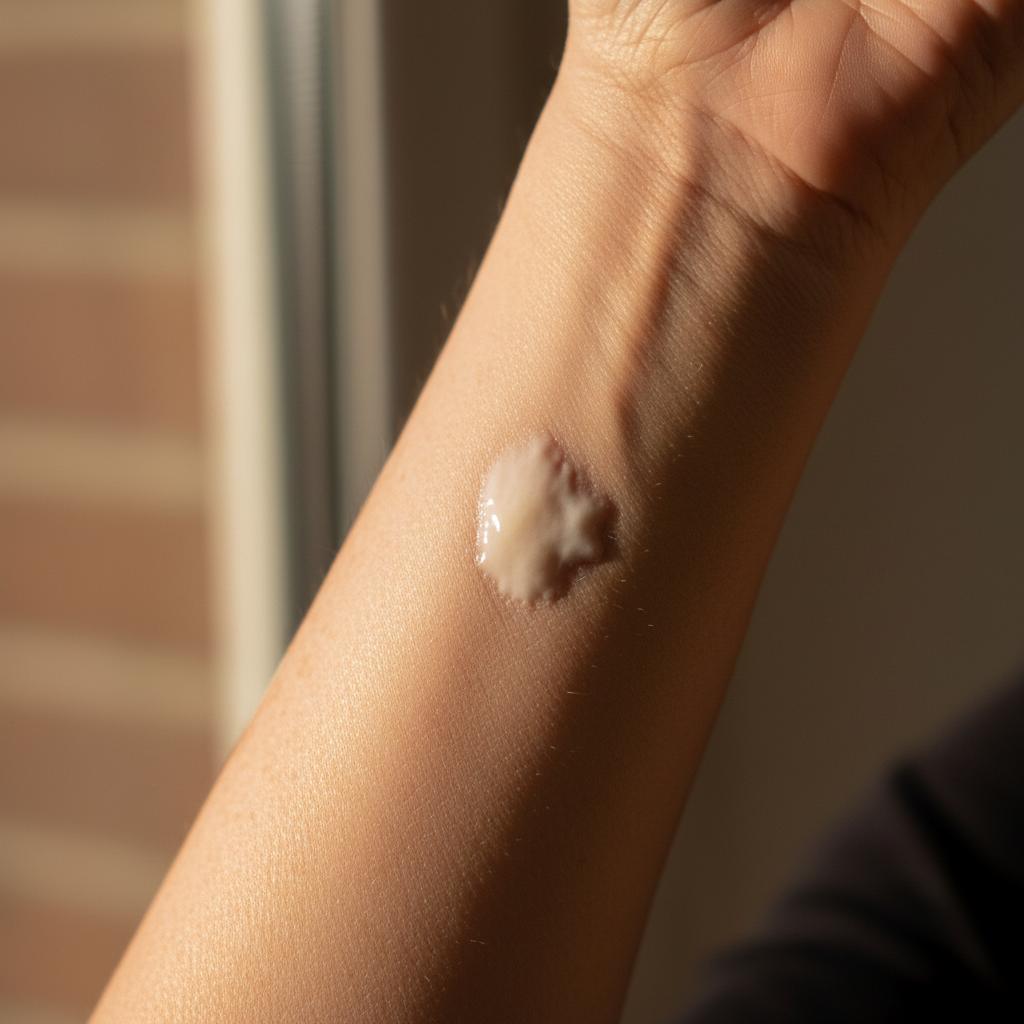
Noticeable changes in your skin’s texture—a sudden patch of tightness, a waxy sheen, or a loss of elasticity—can be unsettling, especially when you are already navigating the transitions of your 40s and 50s. While many women attribute these changes to natural aging or a shift in hormones, localized or systemic skin hardening requires a closer look. Understanding scleroderma vs morphea symptoms in women over 40 is essential for distinguishing between a localized skin condition and a systemic disease that could affect your internal organs.
In this guide, we will explore the nuances of these autoimmune conditions, how they intersect with the hormonal shifts of perimenopause, and why advocacy for the right diagnostic tests is your best tool for long-term health.
What is the difference between systemic scleroderma and morphea?
At their core, both morphea and systemic scleroderma (systemic sclerosis) belong to a family of autoimmune diseases characterized by the overproduction of collagen. However, they are distinct clinical entities with very different trajectories.
Morphea is a localized form of scleroderma. It is primarily confined to the skin and underlying tissues, such as fat, muscle, or bone. It does not affect internal organs or the vascular system. Morphea typically presents as firm, waxy patches (plaques) that may be discolored—often starting with a lilac or reddish border before turning white or yellow in the center. According to the Mayo Clinic, while it can be uncomfortable or cosmetically distressing, it is not life-threatening.
Systemic Scleroderma (Systemic Sclerosis), on the other hand, is a multi-system autoimmune disease. While skin hardening is a hallmark, the disease involves the connective tissue of internal organs, including the lungs, heart, kidneys, and digestive tract. It also involves significant vascular (blood vessel) damage.
For women over 40, distinguishing between the two is vital. While morphea is limited, systemic scleroderma requires aggressive monitoring of organ function. The National Institute of Arthritis and Musculoskeletal and Skin Diseases (NIAMS) notes that systemic sclerosis is significantly more common in women, particularly during their reproductive and menopausal years.
How do localized skin changes differ from systemic autoimmune symptoms?
When you notice a patch of tight skin, your first instinct might be to check if the symptom is "local" or "systemic." The primary differentiator is the presence of symptoms beyond the skin.
In localized morphea, you might see:
- Oval-shaped patches on the torso, arms, or legs.
- A "shiny" appearance to the skin that feels thicker than the surrounding area.
- A loss of hair or sweat glands in the affected patch.
In systemic scleroderma, the skin changes are usually preceded or accompanied by systemic red flags. One of the most common early signs is Raynaud’s phenomenon—where fingers turn white, blue, or red in response to cold or stress. Unlike morphea, systemic sclerosis often causes skin tightening that starts in the fingers (sclerodactyly) and moves up the arms or involves the face.
The following table summarizes the key clinical differences:
| Feature | Morphea (Localized) | Systemic Sclerosis (Scleroderma) |
|---|---|---|
| Primary Site | Skin, fat, and muscle | Skin, blood vessels, and internal organs |
| Raynaud’s Phenomenon | Rarely present | Present in ~95% of cases |
| Organ Involvement | None | Lungs, Heart, Kidneys, GI Tract |
| Skin Pattern | Discrete patches or bands | Symmetrical tightening (fingers, face) |
| Antibody Profile | Usually ANA negative or low titer | Specific antibodies (Scl-70, Centromere) |
If you are experiencing widespread joint pain or extreme fatigue alongside skin changes, you may want to review our perimenopause symptoms checklist to see if your symptoms align with hormonal shifts or if they point toward a more complex autoimmune overlap, such as fibromyalgia-perimenopause symptoms.
Can perimenopause hormone shifts trigger new skin hardening?
The timing of an autoimmune diagnosis often coincides with the late 30s through the 50s, leading researchers to investigate the role of estrogen. Estrogen has a complex relationship with the immune system; it is generally "immunostimulatory," meaning it helps the immune system function, but rapid fluctuations or declines can lead to immune dysregulation.
During perimenopause, the decline in estrogen can lead to:
- Reduced Collagen Quality: While scleroderma involves too much collagen, the decline of estrogen causes a loss of healthy Type I and Type III collagen, making the skin thinner and more prone to injury.
- Increased Inflammation: Estrogen has anti-inflammatory properties. As levels drop, pre-existing autoimmune tendencies may "flare" or become clinically apparent for the first time.
- Vascular Sensitivity: Estrogen helps keep blood vessels flexible. As it declines, symptoms like Raynaud's may worsen, which is a frequent precursor to systemic scleroderma.
For many women, these shifts can also lead to other metabolic changes. If you are noticing skin darkening or "velvety" patches (acanthosis nigricans), it may be worth investigating perimenopause insulin resistance signs, which can sometimes mimic or coexist with autoimmune skin changes.
Why are morphea and scleroderma often misdiagnosed as perimenopause skin aging?
Misdiagnosis is a common hurdle for women in their 40s. Because skin naturally becomes drier, thinner, and less elastic during the menopausal transition, a small patch of morphea or the early stages of systemic skin thickening might be dismissed as "age-related changes" or "just dry skin."
Common reasons for misdiagnosis include:
- Textural Overlap: Estrogen loss leads to skin "crepiness," which can mask the subtle early hardening of scleroderma.
- Symptom Masking: Fatigue and joint pain, which are hallmark signs of systemic scleroderma, are also incredibly common in perimenopause. A physician may attribute your exhaustion to hormonal shifts rather than an underlying autoimmune process.
- Gradual Onset: Scleroderma often develops slowly. By the time the skin changes are obvious, the disease may have been progressing internally for years.
It is also important to remember that autoimmune conditions rarely travel alone. It is not uncommon for women to experience an overlap of conditions. For instance, you might be managing Hashimoto’s and perimenopause overlap, which can cause skin dryness and swelling that complicates a scleroderma diagnosis.
What are the early 'red flag' symptoms of systemic involvement?
If you have been told you have "localized" skin changes, but you suspect something more is happening, you must watch for systemic "red flags." The American College of Rheumatology identifies several symptoms that suggest the disease is moving beyond the skin:
- Raynaud’s Phenomenon: Sudden color changes in the fingers or toes, often accompanied by numbness or pain.
- Puffy Hands: In the "edematous phase," the hands and fingers may look swollen or sausage-like, especially in the morning.
- GERD and Digestive Issues: New or worsening acid reflux, difficulty swallowing, or a "full" feeling after eating very little can indicate that the esophagus is being affected by fibrosis.
- Shortness of Breath: This can be a sign of interstitial lung disease or pulmonary hypertension, two serious complications of systemic scleroderma.
- Telangiectasia: Small, red, mat-like spots on the face, chest, or hands caused by dilated blood vessels.
If you are experiencing these symptoms alongside hormonal changes, discussing HRT for perimenopause beginners guide with your doctor is helpful, but you must also insist on a rheumatology referral to rule out systemic sclerosis.
How does estrogen decline affect skin collagen and autoimmune flares?
Estrogen is a key regulator of skin fibroblasts—the cells responsible for producing collagen and elastin. Research published in the Journal of NIH Research suggests that estrogen helps maintain skin thickness and moisture. When estrogen levels plummet, the balance of collagen production and degradation is disrupted.
In the context of morphea and scleroderma:
- Fibroblast Overactivity: In scleroderma, fibroblasts become hyperactive, pumping out excessive amounts of collagen that "scars" the skin and organs. The loss of estrogen's modulating effect may allow this process to go unchecked in genetically predisposed women.
- Microvascular Damage: Estrogen is cardioprotective and vasoprotective. The decline in estrogen during menopause can exacerbate the vascular damage that defines systemic scleroderma, making Raynaud's symptoms more severe and increasing the risk of digital ulcers.
This is why many women notice their "first" autoimmune symptoms during the transition to menopause. The body’s ability to dampen inflammation is lowered, and the vascular system is under increased stress.
What diagnostic tests distinguish morphea from systemic sclerosis after 40?
If you notice skin changes, a general physical exam is rarely enough. A combination of clinical observation and specific laboratory tests is required to distinguish between localized and systemic disease.
1. Skin Biopsy: A small sample of the affected skin is taken to look for characteristic thickening of the dermis and collagen bundles. While a biopsy can confirm scleroderma-type changes, it cannot always tell if the disease is systemic or localized.
2. Nailfold Capillaroscopy: This is a simple, non-invasive test where a doctor looks at the capillaries at the base of your fingernail under a microscope. According to the Cleveland Clinic, patients with systemic scleroderma will often have enlarged or missing capillaries, whereas those with morphea will typically have normal nailfold patterns.
3. Blood Work (The Scleroderma Panel):
- ANA (Antinuclear Antibody): A positive ANA is found in nearly all systemic scleroderma patients, though it can also be positive in other conditions like Hashimoto's.
- Centromere Antibody: Highly associated with Limited Cutaneous Systemic Sclerosis (formerly CREST syndrome).
- Scl-70 (Topoisomerase I) Antibody: Associated with Diffuse Cutaneous Systemic Sclerosis and a higher risk of lung involvement.
- RNA Polymerase III: Associated with rapid skin involvement and a higher risk of kidney issues.
4. Pulmonary Function Tests (PFTs) and Imaging: If systemic involvement is suspected, doctors will use PFTs or a high-resolution CT scan to check for lung fibrosis, even if you don't feel short of breath yet.
Understanding these tests is the first step in self-advocacy. If your doctor dismisses your skin patches as "just aging," ask specifically for a nailfold capillaroscopy or an ANA screen to ensure nothing is being missed during this pivotal time in your health journey. By identifying the difference between scleroderma vs morphea early, you can take control of your treatment and protect your long-term vitality. High-quality care starts with accurate identification—don't let your symptoms be "written off" as just another part of getting older.
FAQ
Common questions
Can morphea turn into systemic scleroderma?
Morphea is limited to patches of skin and does not affect internal organs or shorten life expectancy. Systemic scleroderma involves the skin, blood vessels, and internal organs (like lungs and kidneys) and requires lifelong specialist management.
Is morphea an autoimmune disease?
No. While they are both types of scleroderma, they are considered distinct conditions. It is extremely rare for a person diagnosed with localized morphea to later develop systemic sclerosis, though both involve similar collagen-production issues.
What does early systemic scleroderma feel like?
Yes, morphea is considered a localized autoimmune skin disorder where the immune system mistakenly attacks skin cells, leading to excessive collagen production and hardening.
Why is scleroderma more common in women over 40?
Early signs often include Raynaud’s phenomenon (fingers turning white/blue in the cold), puffy or swollen 'sausage' fingers, unexplained fatigue, and persistent heartburn or acid reflux.
Can estrogen therapy help with scleroderma skin changes?
The peak onset for systemic scleroderma is between ages 35 and 55. Researchers believe the intersection of genetic predisposition and the significant hormonal shifts (like estrogen decline) during these years may trigger the condition.
How do I know if my skin changes are just menopause aging?
While HRT helps with general skin aging and elasticity, it is not a primary treatment for scleroderma. However, maintaining hormonal balance may help manage some overlapping perimenopausal symptoms that exacerbate autoimmune fatigue.
Want this in your inbox each Sunday?
New articles, the science you can actually use, and the occasional rant.
Keep reading

Perimenopause Symptoms Checklist: 38 Signs You're Not Imagining
A comprehensive, doctor-reviewed checklist of perimenopause symptoms — from the obvious hot flashes to the weird ones nobody warned you about.
9 min read

Hashimoto's and Perimenopause: Why Symptoms Overlap (and How to Tell Them Apart)
Fatigue, brain fog, weight gain, hair loss — Hashimoto's and perimenopause share most symptoms. Here's how to tell what's flaring, what's hormonal, and what to ask your doctor.
11 min read

Fibromyalgia and Perimenopause: When Two Storms Collide
Why fibromyalgia almost always flares during perimenopause, what changes in your nervous system, and the small daily shifts that actually help.
10 min read